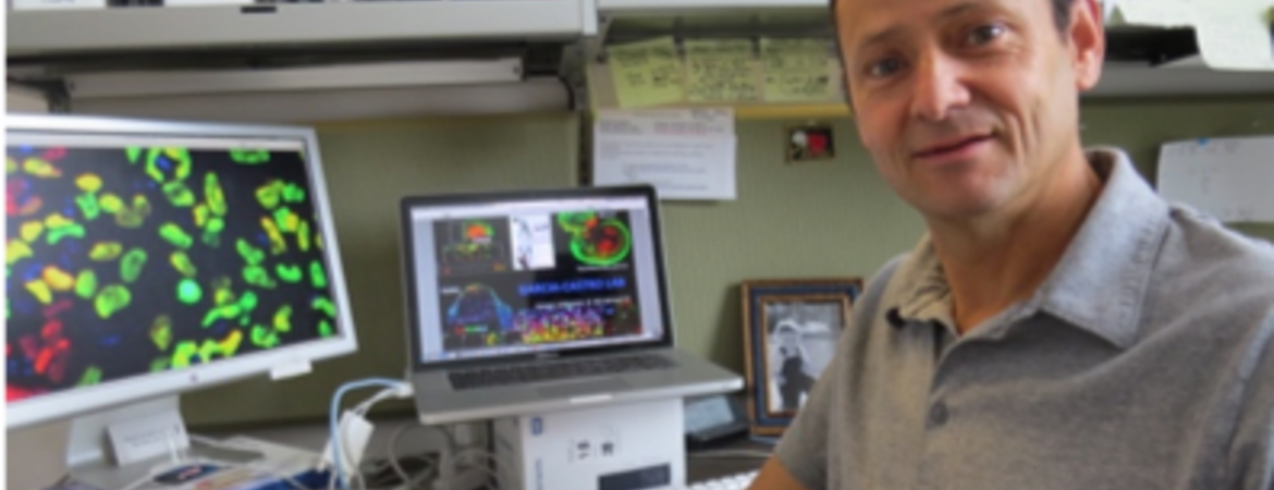
Neural crest cells arise early in the development of vertebrates, migrate extensively through the embryo, and differentiate to give rise to a wide array of derivatives. The proper functioning of these embryonic cells is critical for human development and health. When neural crest biology fails, various birth defects and illnesses, such as cleft lip or palate, Waardenburg syndrome, and melanoma, can result.
In 2016, Martín García-Castro's lab reported a fast and robust model for developing neural crest cells. The lab has now published two research papers on these cells.
In the paper, “Human neural crest induction by temporal modulation of WNT activation,” published in Developmental Biology, the authors report that a two-day pulse of “WNT” exposure (WNT is an important cell signaling molecule) is sufficient to trigger robust formation of neural crest cells across a wide range of culture size, enabling a high yield of cells.
“The generation of these cells in culture dishes holds a great promise for clinical uses and basic biology research alike,” said García-Castro, an associate professor of biomedical sciences in the School of Medicine.
In the second paper, “FGF modulates the axial identity of trunk hPSC-derived neural crest but not the cranial-trunk decision,” published in Stem Cell Reports, the authors further improve the efficiency of neural crest generation in culture leading to an impressive 90% yield across multiple cell lines. The lab achieved this by combining the two-day pulse of WNT with a method controlling the variability of another important signal, bone morphogenetic protein.
“By providing animal product-free culture conditions this protocol brings us closer to the use of these cells in the clinic,” García-Castro said.
